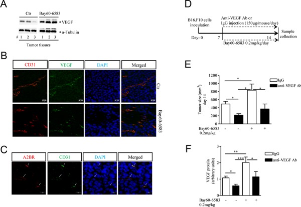
Figure 2. Increased tumor angiogenesis in melanoma-bearing mice treated with Bay60-6583 compared with control mice.

A. Western blotting analysis of VEGF protein expression in melanoma tissue lysates harvested from mice treated with Bay60-6583 0.2 mk/kg or vehicle (ctr). B. Immunofluorescence staining of CD31 and VEGF double-positive vessels in melanoma tissue sections of mice treated with Bay60-6583 or vehicle are shown. Magnification 63x. Scale bars represent 20 μm. C. Immunofluorescence staining of CD31 and A2B receptor in melanoma sections is shown. Magnification 63x. Scale bars represent 20 μm. D. C57Bl6j mice implanted with B16.F10 melanoma cells (day 0), treated from day 7 with Bay60-6583 (0.2 mg/kg p.t.) or vehicle, were injected with anti-VEGF antibody (150 μg/mouse i.p.) or IgG control. At day 14 mice were sacrificed to collect tumor tissues for further analyses. E. Tumor volume measured at day 14 after tumor cell implantation of mice treated with Bay60-6583 or vehicle receiving anti-VEGF antibody or IgG as above. F. Analysis of VEGF protein expression in melanoma tissue harvested from mice treated as described above. Data are from three independent experiments and represent mean ± SEM (n = 6–10 per group). *p < 0.05, **p < 0.01 and ***p < 0.001.
