
Figure 7. DECA-14 inhibits migratory properties of macrophages in vitro and in vivo.
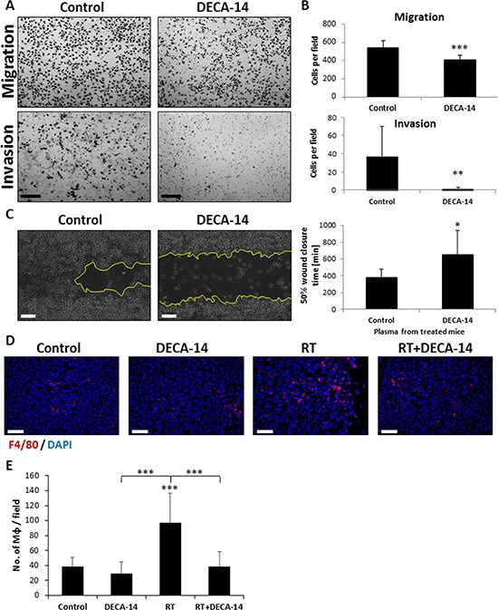
A-B. The invasion and migration properties of J774 macrophages were assessed in the presence or absence of DECA-14 (100 nM) using the Boyden chamber assay. Images of the membranes were captured (A) and the number of cells per field was counted (B). Scale bars = 200 μm. C. A scratch wound assay was performed on J774 cells cultured in the presence of plasma obtained from mice 24 hours after they were treated with DECA-14, or from control mice. The percentage of wound closure time was evaluated and plotted using ImagePro Premier software. Representative images of 600 min time-point are presented. D–E. Eight-to-ten week old SCID mice were orthotopically implanted with SW480 tumors. After 4 weeks, the mice were intraperitoneally injected with 2.5 mg/kg DECA-14, and 24 hours later mice were either irradiated in the tumor area (RT) or left untreated (Control)(n = 5–6 mice/group). Three days later, tumors were harvested and (D) immunostained for macrophages (F4/80, red). Nuclei were stained with DAPI (blue). Scale bars = 100 μm. (E) The number of macrophages per field were counted (n > 10 fields/group). *0.05 > p > 0.01; **0.01 > p > 0.001; ***p < 0.001.