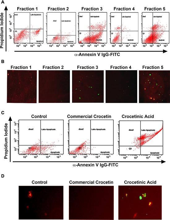
Figure 2. Crocetinic acid induces apoptosis.

A, B. MiaPaCa-2 cells were incubated with 1 μM of the various fraction and levels of apoptosis was determined by flow cytometry (A) and fluorescence microscopy (B). In flow cytometry, propidium iodide staining is for dead cells (Y-axis), while annexin V was determined with a specific antibody conjugated to fluorescein isothiocyanates (FITC). For immunofluorescence, cells treated with FITC were imaged under a fluorescent microscope. Fraction 3 and 5 showed the maximum level of apoptosis by both methods. C, D. Panc-1 cells were incubated with 1 μM purified crocetinic acid and commercial crocetin and again tested for apoptosis by flow cytometry and immunofluorescence. Again, there was significantly higher numbers of apoptotic cells in crocetinic acid treated cells.
