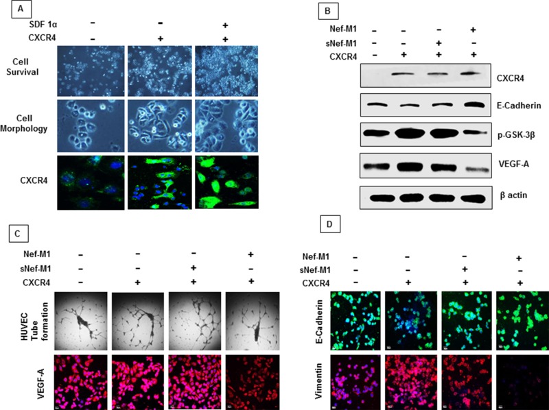
Figure 6. A. Proliferation and morphologic features of MDA-MB468 cells overexpressing CXCR4.
Cell morphologic changes are shown in phase-contrast images. The CXCR4 and its ligand SDF-1α induces proliferation and morphological changes associated with tumor progression. SDF-1α induces internalization of CXCR4. B. Cell lysates from MDA-MB468 control or CXCR4-expressing cells were immunoblotted with antibodies for anti-CXCR4, E-cadherin, p-GSK-3β, VEGF-A or β-actin. Cells that express CXCR4 became susceptible to Nef-M1 peptide induced inhibition of p-GSK-3β expression (mesenchymal signature), VEGF-A expression (tumor angiogenesis) and induced elevation of E cadherin (epithelial signature). C. Cells that express CXCR4 became susceptible to Nef-M1 peptide induced inhibition of capillary network formation and VEGF-A expression as detected by tube formation assay and immunofluorescence respectively. D. Cells that express CXCR4 became susceptible to Nef-M1 peptide induced elevation of E cadherin (epithelial signature) and inhibition of vimentin expression (mesenchymal signature) as detected by immunofluorescence analysis.

