
Figure 1. HOTAIR and SETD2 expression in human liver cancer tissue.
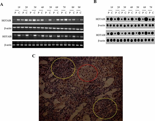
A. RT-PCR analysis of HOTAIR in liver cancer tissue (C) and its paracancerous liver tissues (P) respectively (indicated in upper). β-actin as internal control. B. Nuclear Run on analysis of HOTAIR in liver cancer tissue (C) and its paracancerous liver tissues (P) respectively (indicated in upper). β-actin as internal control. C. The representative analytic results of SETD2 immunohistochemistry staining of formalin-fixed, paraffin-embedded human liver cancer tissue (indicated with yellow Dotted circles) and their paired adjacent noncancerous tissues (indicated with Dotted red circles) from the same patient. (DAB stainning, original magnification × 100).