
Figure 8. The rescued experiment of carcinogenesis effect of the HOTAIR.
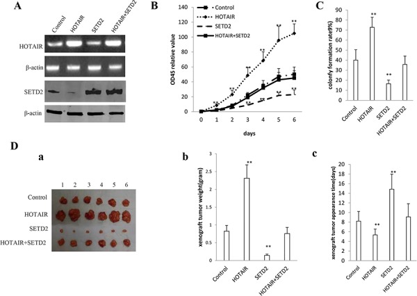
SETD2 overexpression abrogated the HOTAIR oncogenic function in stable human liver cancer stem cell (hLCSC) transfected with pCMV6-A-GFP, pCMV6-A-GFP-HOTAIR, pcDNA3.1-SETD2, pCMV6-A-GFP-HOTAIR plus pcDNA3.1-SETD2. A. The RT-PCR analysis of HOTAIR (upper) and the western blotting analysis with anti-SETD2 (lower). β -actin as internal control. B. Cells growth assay using CCK8. Each value was presented as mean ± standard error of the mean (SEM). C. Cells colony-formation efficiency assay. Each value was presented as mean ± standard error of the mean (SEM). D. In vivo test in human liver cancer stem cells (hLCSC) transfected with pCMV6-A-GFP, pCMV6-A-GFP-HOTAIR, pcDNA3.1-SETD2, pCMV6-A-GFP-HOTAIR plus pcDNA3.1-SETD2. a. The mice were stratified and the tumors were recovered. The photography of xenograft tumor in the three groups (indicated in left). b. The wet weight of each tumor was determined for each mouse. Each value was presented as mean ± standard error of the mean (SEM). c. The xenograft appearance time (day). Each value was presented as mean ± standard error of the mean (SEM).