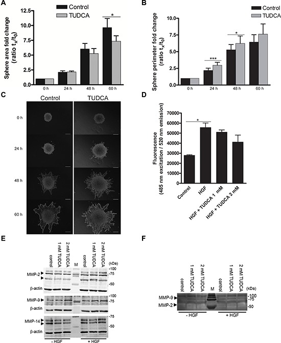
Figure 5. Effects of TUDCA on HCC cell invasion.

A. The area and B. length of the outside boundary (perimeter) of collagen-embedded Hepa1-6 spheroids (n = 9) was measured at 0, 24, 48 and 60 h of incubation with 2 mM TUDCA or control medium. Results are representative of 2 independent experiments. C. Representative images of invasive capacity of cells present in a collagen-embedded multicellular spheroid are shown at different time points. The multicellular spheroids are approx. 150 μm in diameter at start. At 60 h, the spheroid perimeter is marked. Scale bar is 50 μm. D. Boyden chamber invasion assay with Hepa1-6 cells following 48 h of incubation with control medium, control medium with chemoattractant hepatocyte growth factor (HGF) without or with 1–2 mM TUDCA. Samples were run in quadruplicate. E. Western blot analysis of MMP-2, -9 and -14 levels in Hepa1-6 cell lysates of control cells and cells treated with 1 or 2 mM TUDCA (48 h), in the presence or absence of 50 ng/ml HGF stimulation. Arrows indicate MMP-2 (72 and 63–66 kDa), MMP-9 (78–82 kDa) and MMP-14 (66–57 kDa). The β-actin signal is used as loading control. F. Gelatin zymography of concentrated culture medium of Hepa1-6 control cells or cells treated with 1 or 2 mM TUDCA (48 h), in the presence or absence of 50 ng/ml HGF stimulation. The white bands indicate MMP-activity; arrowheads: signal for MMP-9 (glycosylated, 92 kDa) and MMP-2 (58/62 kDa, active). Values represent the mean ± SD. *p < 0.05, ***p < 0.001.
