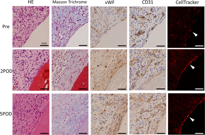
Fig 6. Histological examination of the luminal side of the vascular graft.
At pre-implantation, the vascular endothelial cells distribute to the entire area of the graft. Conversely, after implantation, vWF, CD31 and CellTracker Red-positive endothelial cells are seen at the inner lumen of the vessel. Furthermore, the vascular endothelial cells cover the inner surface of the vessel more continuously on the fifth day than on the second day.

