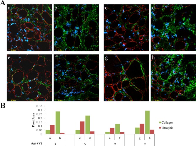
Figure 1. Utrophin and collagen type I levels in muscles of DMD patients.

QF biopsies taken from DMD patients were double-immunostained with utrophin (red) and collagen type I (green) antibodies, nuclei were stained with DAPI (blue), and all were visualized by confocal microscopy. A. Depiction of areas rich in either utrophin or collagen type I that were taken from four DMD patients. B. Image analysis quantification of utrophin and collagen type I levels presented as pixels/unit area. The levels of the collagen and utrophin signals were evaluated with NIS-Elements microscope imaging software (Nikon Instruments Inc). a, b – 3-year-old DMD patient; c, d – 5-year-old DMD patient; e, f and g, h – two 9-year-old DMD patients. Note that regardless of age, areas that are rich in collagen type I exhibit low levels of utrophin and vice versa.
