
Figure 2. Z-stacks photo images of muscle biopsies of DMD patients.
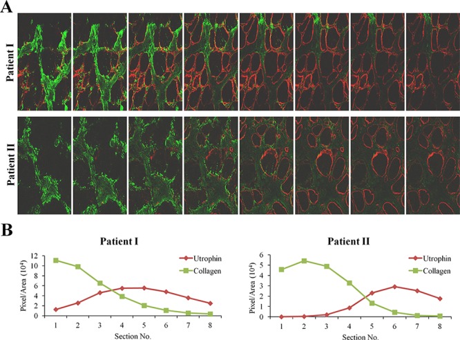
A. QF biopsies from two patients (patient I - 4 years; patient II - 8 years) were double-immunostained with utrophin (red) and collagen type I (green) antibodies. Utrophin and collagen type I were visualized by confocal microscopy, and Z-stacks photographs (taken at 1.2 μm intervals, extending to 8-μm depth in the dystrophic muscle) were taken. B. Image analysis quantification of utrophin and collagen type I levels presented as pixels/unit area.