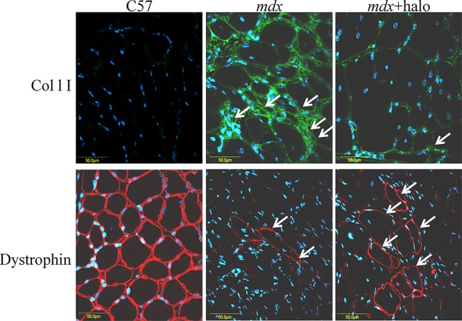
Figure 6. Inhibition of fibrosis and numbers of RFs.

Diaphragms of wild-type C57 mice, of untreated mdx mice, and of mdx mice treated with halofuginone at 7.5 μg/mouse for 10 weeks starting from 4 weeks of age, were immunostained with dystrophin antibodies (red) and collagen type I antibodies (green), nuclei were stained with DAPI (blue) and all were visualized by confocal microscopy. In the diaphragm of the C57/BL mice no collagen was observed and all diaphragm myofibers were stained with dystrophin antibodies. In the mdx diaphragm high levels of collagen were observed, together with some RFs. A major decrease in collagen type I levels and increase in the number of RFs were observed after halofuginone treatment.
