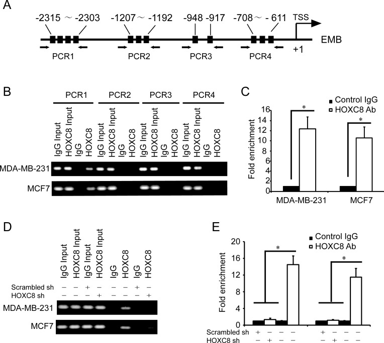
Figure 3. HOXC8 binds to the promoter of EMB.
A. A schematic diagram for the positions of putative HOX protein binding sites in the promoter of EMB. Arrows show the regions for PCR primers amplification. B. ChIP was performed on MDA-MB-231 or MCF7 cells using HOXC8 antibody or mouse IgG and the immunoprecipitated chromatin DNA was subjected to PCR using primers to amplify the region of EMB promoter. C. ChIP was performed on MDA-MB-231 or MCF7 cells using HOXC8 antibody or IgG as control, and precipitated chromatin DNA were subjected to qPCR using primers to amplify the regions of EMB promoter. D. MDA-MB-231 and MCF7 cells were lentivirally transduced with HOXC8 shRNA vectors or scrambled shRNA vectors, and HOXC8 ChIP assays were performed with IgG as negative control. E. MDA-MB-231 and MCF7 cells were lentivirally transduced with HOXC8 shRNA vectors or scrambled shRNA vectors, and HOXC8 ChIP assays were performed with IgG as negative control. Precipitated chromatin DNA were subjected to qPCR using primers to amplify the regions of EMB promoter.

