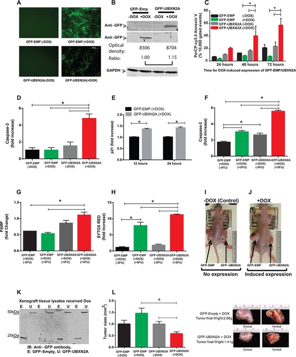
Figure 2. Induction of UBXN2A slows the growth of a colon cancer tumor ex vivo and in a mouse xenograft model by 50%.

A. A Tet-on regulated inducible HCT-116 colon cancer cell line was established successfully. 48 hours incubation with Doxycycline (DOX, 10 mg/ml) induces an equal expression of GFP-empty (EMP) or GFP-UBXN2A in HCT-116 cells. Images were taken using an inverted fluorescent microscope. B. WB showing the expression of GFP-empty and GFP-UBXN2A proteins in HCT-116 cells when treated with DOX for 48 hours. An anti-GFP antibody was used to detect the levels of GFP alone or of GFP-UBXN2A fusion proteins, while GAPDH was used as a loading control. Optical density of bands were measured by Image Studio software C. Inducible cells (GFP-EMP or GFP-UBXN2A) were treated with DOX for the indicated times. Cells were then stained with PerCP-Cy5.5 Annexin V antibody, and a total of 10, 000 gated events were analyzed by flow cytometry. DOX-induced UBXN2A expression in 48 and 72 hours showed a significant increase in annexin binding in those cells. The data is shown as mean ± SEM of three independent experiments in trplicate (n = 3) where *p < 0.001 using Tukey's modified student's t-test. D. Inducible cells (GFP-EMP or GFP-UBXN2A) were treated with DOX for 48 hours, followed by staining with Alexa Fluor 546 anti-caspase-3 antibody. Cells were analyzed using flow cytometry and collected data was normalized with GFP-EMP (+DOX). The DOX-induced UBXN2A expression significantly increased caspase-3 levels (n = 3, *p < 0.05). E. Inducible cells (GFP-EMP or GFP-UBXN2A) were treated with DOX for the indicated times. Immunofluoresence of p21 was performed. Intensity of fluorochrome signals from a confocal microscope were analyzed by the Image J (NIH, Bethesda, MD) program and plotted. The data was normalized with GFP-EMP (+DOX). The expression of UBXN2A significantly increased levels of p21 (n = 100, *p < 0.001). F–G. HCT-116 cells expressing GFP-EMP and GFP-UBXN2A under DOX were treated with 5-FU (100 μM) for 48 hours. Cells were then stained with Alexa Fluor 546 anti-caspase-3 (F) or anti-cleaved PARP (G) antibodies, followed by flow cytometry analysis. The induced UBXN2A significantly enhanced 5-FU-induced increase in caspase-3 and cleaved PARP levels (n = 3, *p < 0.05). H. Inducible HCT-116 cells were incubated with DOX and 5-FU for 48 hours, stained with Sytox Red, and then the population of dead cells was analyzed using flow cytometry. UBXN2A significantly enhanced 5-FU-induced cell death (n = 3, *p < 0.05). I–J. 1 × 10⁷ Tet-on inducible cells were injected subcutaneously into the lower flanks of nude mice. The animals with palpable tumors (< 5 mm3) for early-stage tumor response were divided into two groups after injection to be fed with a standard diet (controls) or a DOX-containing diet (625 mg/kg). Tumor volumes in mm3 were determined with digital calipers by the formula Volume = (width) 2 × length/2 every second day for 40 days. K. Expression of GFP-empty or GFP-UBXN2A proteins in dissected xenografts confirmed successful tumor responses to DOX after 40 days. L. Growth of tumors with and without DOX on day 40. Treatment with DOX significantly decreases tumor size and weight by more than 50%. The data is shown as mean ± SEM of 9 mice (n = 4 for control and n = 5 for DOX) where *p < 0.05 using Tukey's modified student's t-test.
