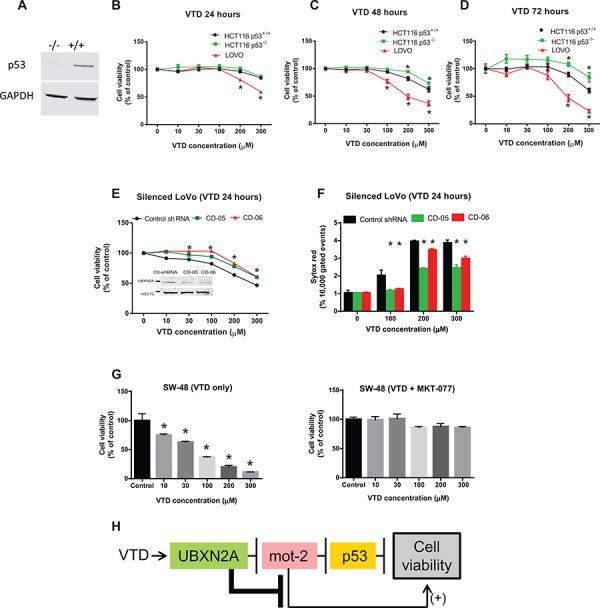
Figure 5. VTD functions via the UBXN2A-mot-2-p53 axis.

A. WB confirmed the presence and the absence of p53 protein in HCT-116 p53(+/+) and HCT-116 p53(−/−) cancer cell lines, respectively. B–D. The effect of VTD on the viability of poorly-differentiated (HCT-116 p53(+/+) and HCT-116 p53 (−/−)) and well-differentiated (LoVo) colon cancer cell lines was determined using an MTT assay. VTD's cytotoxic effect on all three cell lines was dose dependent in (B) 24 hours, (C) 48 hours, and (D) 72 hours. As compared to the HCT-116 p53(+/+) cells, HCT-116 p53(−/−) cells showed more resistance to VTD's effects, whereas well-differentiated (LoVo) colon cancer cells showed the highest sensitivity to VTD. E. LoVo cells were stably silenced for UBXN2A using a UBXN2A-shRNA along with a scrambled shRNA. Viability of UBXN2A-silenced cells was found to be significantly higher as compared to control cells upon treatment with VTD. F. In another set of experiments, UBXN2A-silenced cells were treated with VTD (100, 200, and 300 μM) for 24 hours. Cells were then labelled with Sytox Red followed by flow cytometry analysis. Results showed silencing of UBXN2A significantly decreases cell death in response to VTD. G. SW48 well-differentiated colon cancer cells with WT-p53 were treated with MKT-077 (5 μg/ml), a mot-2 inhibitor, along with VTD for 72 hours. A clonogenic survival assay showed VTD signifcantly decreases the colony number of SW-48. However, preincubation with MKT-077 neutralizes the cytotoxic effect of VTD. The data is shown as mean ± SEM of three independent experiments (n = 3) in triplicate where *p < 0.05 using Bonferroni's modified student's t-test. H. This flowchart recapped the sequence of proteins which activate and function upon VTD exposure to decrease cell viability.
