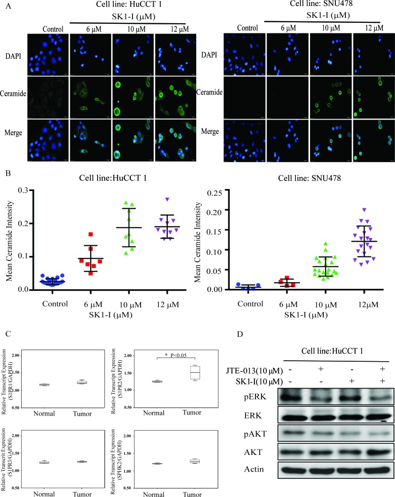
Figure 4. SK1-I increased intracellular ceramide and inhibited ERK and AKT signaling when combined with JTE-013.
A. HuCCT1 and SNU478 were treated with SK1-I 0 μM, 6 μM, 10 μM, and 12 μM for 48 h. Cells were fixed and stained with anti-ceramide (green) and DAPI (blue) and then imaged by fluorescence microscopy. B. The intensity of ceramide expression was greater in HuCCT1 and SNU478 cells treated with SK1-I (6 μM, 10 μM, and 12 μM) than in the controls. C. The mRNA expression of S1PR2 was higher in CCA than in normal liver tissue, as determined by qRT-PCR (p < 0.05). D. Western blot analysis of phosphorylated ERK and AKT after 15 min of treatment with SK1-I 10 μM and JTE-013 10 μM without serum.

