
Figure 3. DIM inhibited the adhesion, migration and invasion of SMMC-7721 and MHCC-97H.
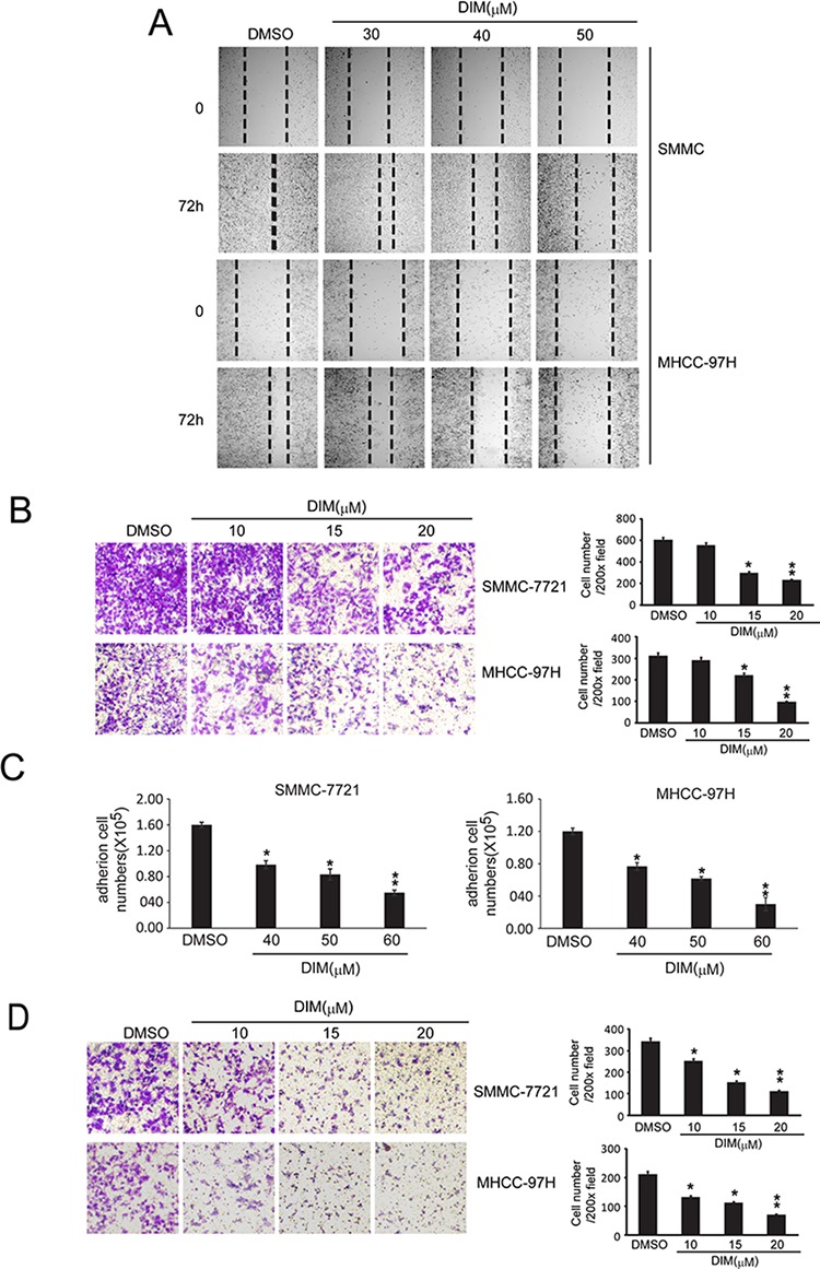
A. Wound healing assay were conducted. SMMC-7721 and MHCC-97H cells were seeded into 6-well plates and treated with DMSO or DIM (30 h, 40 h and 50 μM for 72 hs). Photographs were taken after treatment. B. Migration tests were performed by transwells inserts without basement membrane extract. SMMC-7721 and MHCC-97H (1 × 10⁵ cells per well) were seeded in insert with 200 μl no-serum medium containing different concentration of DIM (5, 10 and 15 μM), 800 μl medium containing 5% FBS was added in bottom wells and cells were incubated for 16 hours. Cells were stained with Giemsa. C. Cell adhesion assay. SMMC-7721 and MHCC-97H were treated in 6-well culture dishes with DIM at 30, 40 and 50 μM for 48 hours in medium with 10% FBS. After that, Cells (5 × 10⁵ cells/well) were plated in 6-well culture dishes and allowed to adhere for 2.5 h. After that, adhered cells were counted after staining with 0.4% trypan blue solution. D. Cells invasion assay was performed by transwell inserts with basement membrane extract. SMMC-7721 and MHCC-97H (1 × 10⁵ cells per well) were seeded in inserts with 200 μl no-serum medium containing different concentration of DIM (10, 15 and 20 μM), 800 μl medium containing 5% FBS was added in bottom wells and cells were incubated for 24 h. Cells were stained with Giemsa. Values represent mean ± SD of three independent experiments. *P < 0.05, **P < 0.01, ***P < 0.001 compared with the untreated control (dose 0).