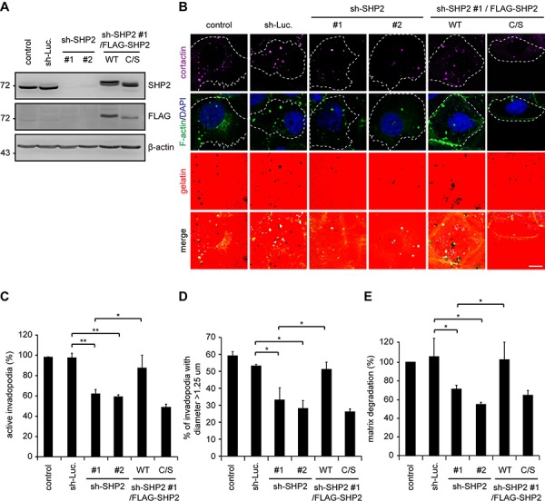
Figure 2. The suppression of invadopodia formation by SHP2 depletion is restored by re-expression of SHP2 but not its catalytically defective mutant in SAS cells.

A. shRNAs specific to luciferase (sh-Luc.) or SHP2 (sh-SHP2; clones #1 and #2) were stably expressed in SAS cells. Wild-type human FLAG-tagged SHP2 (FLAG-SHP2 WT) or the C459S (C/S) mutant, which is deficient in phosphatase activity, were re-expressed in the cells expressing sh-SHP2 #1. A equal amounts of whole cell lysates was analyzed by immunoblotting with the indicated antibodies. B. The cells were seeded on Alexa Fluor 546-conjugated gelatin-coated coverslips for 54 h. The cells were fixed and then stained for F-actin, cortactin and DAPI. Active invadopodia were defined by colocalization of F-actin and cortactin with degraded gelatin. Scale bar, 10 μm. C. Quantitation of active invadopodia formation in B. The number of active invadopodia per cell was determined (n > 150). The data are expressed as the percentage relative to the control SAS cells, which was set 100%. Values (means ± s.d.) are based on three independent experiments; *P < 0.05; **P < 0.001. D. Quantitation of the diameter of active invadopodia in B. The percentage of active invadopodia with a diameter over 1.25 μm out of the total number of active invadopodia (n > 100) is shown. Values (means ± s.d.) are based on three independent experiments; *P < 0.05; **P < 0.001. E. Quantitative results for the matrix degradation assay. The data are expressed as the percentage relative to the level of the control SAS cells, which was set 100%. Values (means ± s.d.) are from three independent experiments; *P < 0.05; **P < 0.001.
