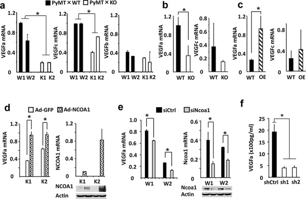
Figure 3. NCOA1 regulates VEGFa expression in breast tumor cells.

a. Relative expression levels of VEGFa, VEGFc and VEGFb mRNAs in W1, W2, K1 and K2 cells measured by QPCR. b. Relative expression levels of VEGFa and VEGFc mRNAs in Tg(MMTV-PyMT) (WT) and Tg(MMTV-PyMT) × Ncoa1−/− (KO) mouse mammary tumors (n = 5) measured by QPCR. c. Relative expression levels of VEGFa and VEGFc mRNA levels in Tg(MMTV-TVA/RCAS-PyMT) (WT) and Tg(MMTV-TVA/RCAS-PyMT) × Tg(MMTV-NCOA1) (OE) mouse mammary tumors (n = 10) measured by QPCR. d. Relative expression levels of VEGFa mRNA in K1 and K2 cells with adenovirus-mediated GFP or NCOA1 expression (left panel). The expression levels of NCOA1 were analyzed by both QPCR and Western blotting (right panel). As expected, human NCOA1 mRNA was not expressed in the mouse K1 and K2 cells. e. Relative expression levels of VEGFa and Ncoa1 mRNAs in W1 and W2 cells transfected with siCtrl or Ncoa1 siRNAs as indicated. The measurement was carried out by QPCR. Ncoa1 knockdown efficiency was also analyzed by Western blotting. f. Secreted VEGFa concentrations in the conditioned media of MDA-MB-231 cells expressing non-targeting control shRNA (shCtrl) or NCOA1 mRNA-targeting shRNAs (sh1 and sh2). NCOA1 knockdown efficiency in these cells was shown in Figure 2e. The * in all panels indicates p < 0.05 by Student's t test.
