
Figure 5. NCOA1 cooperates with HIF1α and AP-1 to activate the VEGFa promoter.
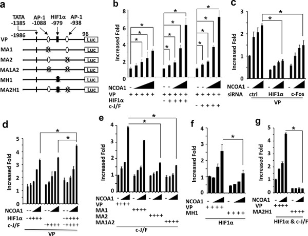
a. Wild type and mutant VEGFa promoter-reporter constructs. The locations are labeled by setting the transcriptional starting site as bp 1. TATA box and AP-1 and HIF1α binding sites are indicated. VP, wild type VEGFa promoter; MA1 or MA2, mutant VEGFa promoters with deletion of the first or second AP-1 site; MA1A2, mutant VEGFa promoter with deletion of both AP-1 sites; MH1, mutant VEGFa promoter with deletion of the HIF1α binding site; MA2H1, mutant VEGFa promoter with deletion of the HIF1α and the second AP-1 binding sites; Luc, luciferase. b. Enhancement of VEGFa promoter activity by NCOA1 alone or NCOA1 with HIF1α or c-Jun/c-Fos (c-J/F). HeLa cells in 24-well plates were co-transfected with 200 ng of VP-Luc plasmid and 0, 150, 300, 600, 900 or 1200 ng of NCOA1 expression plasmid (left panel) or 0, 0, 150, 300 and 600 ng of NCOA1 expression plasmid with 100 ng of HIF1α expression plasmid (middle panel) or c-J/F (50 ng each, right panel) expression plasmids as indicated. c. Knockdown of HIF1α or c-Fos reduced NCOA1-promoted activity of the VEGFa promoter. HeLa cells were transfected with 0, 150, 300 and 600 ng of NCOA1 expression plasmid and 200 ng of VP-Luc reporter plasmid. d. NCOA1 promotes HIF1α and c-J/F mediated activation of the VEGFα promoter. HeLa cells were co-transfected with VP-Luc reporter, NCOA1 and HIF1α, c-Jun/c-Fos or both HIF1α and c-Jun/c-Fos as described above for panel c. e–f. Deletion of the second AP-1 site or the HIF1α binding site reduced NCOA1/C-J/F or NCOA1/HIF1α-promoted activity of the VEGFa promoter. HeLa cells in 24-well plates were co-transfected with same amounts of NCOA1 plasmid as that in panel c, 100 ng of c-J/F plasmids or HIF1α plasmid, and 200 ng of VP-Luc, MA1-Luc, MA2-Luc or MA1A2-Luc reporter plasmid as indicated. g. Deletion of both the second AP-1 and the HIF1α binding sites diminishes NCOA1-enhanced HIF1α and C-J/F-mediated activation of the VEGFa promoter. HeLa cells were transfected with the indicated plasmids as described above for panel f. In all experiments, luciferase activity was assayed 48 hours after transfection and normalized to the total protein amount assayed for each sample. All experiments were repeated at least three times. The * in all panels indicates p < 0.05 by One-Way ANOVA test.