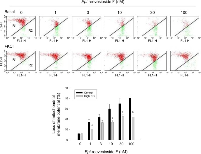
Figure 6. Effect of Epi-reevesioside F on mitochondrial membrane potential (ΔΨm).
T98 cells were incubated in the absence or presence of Epi-reevesioside F for 24 hours with or without extracellular potassium supplements (10.7 mM). After the treatment, the cells were incubated with JC-1 for the detection of ΔΨm using flow cytometric analysis. Data are expressed as mean±SEM of four independent determinations. *P < 0.05 and **P < 0.01 compared with Epi-reevesioside F alone.

