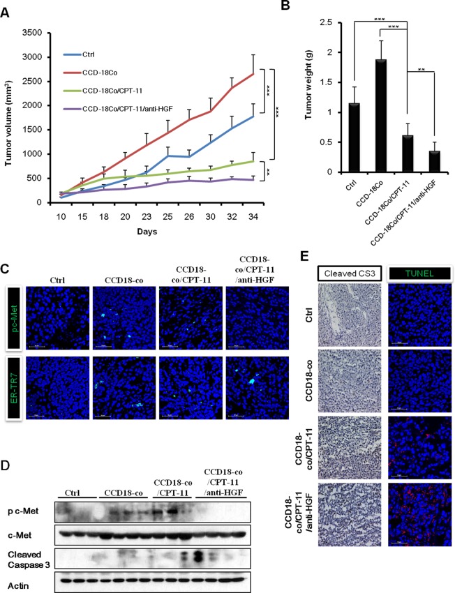
Figure 5. Combination of humanized anti-HGF antibody and CPT-11 inhibits tumor growth in vivo HCT-116 cells with or without CCD-18co cells were inoculated subcutaneously into NOD/SCID mice.
Mice in the control group of received PBS. Mice in the treatment groups received CPT-11 (10 mg/kg; 3 times/wk; intraperitoneally) with or without humanized anti-HGF antibody (10 mg/kg; 2 times/wk; intraperitoneally). A. Tumor volume was measured every 3 or 4 days and calculated. B. Average tumor weights were measured on day 34. The differences were statically significant between the combination treatments versus single treatment. Significant differences were evaluated using an unpaired two-tailed Student's t-test. (Error bars denote the standard deviation [SD]) (**p < 0.001 and ***p < 0.001). All four treatment groups were well tolerated, as total mouse body weights remained unchanged (<10 % fluctuation). C. Immunofluoresence images of phospho-c-Met receptors and a fibroblast marker (ER-TR7) in tumors. Co-staining with DAPI was done to show nuclei. Frozen tumor specimens were subjected to immunofluorescence analysis using anti-phospho-c-Met antibody (green; upper panel) or anti-ER-TR7 antibody (green; lower panel). D. Western blot analysis for cleaved caspases-3 and phospho-c-Met receptors. E. Immunohistochemistry and TUNEL assays were performed to observe apoptotic cell death in tumors. Immunohistochemical analysis in tumors was performed using an antibody against cleaved caspase-3 (left panel). Tumor sections were stained with hematoxylin for counterstaining. The red fluorescent staining indicates positive apoptotic cells and DAPI staining was used as a nuclear counterstaining (right panel).

