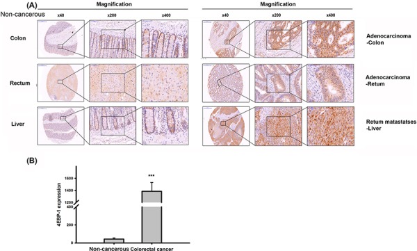
Figure 2. Immnuohistochemical staining of 4E-BP1 in a tissue microarray.

A. Representive 4E-BP1 staining of CRC specimens and corresponding non-cancerous tissues. B. The statisitc result of 4E-BP1 expression in CRC and paired normal specimens by Image J. ***P < 0.001.
