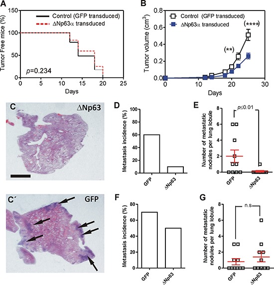
Figure 3. Expression of huΔNp63α decreases spontaneous metastasis development.

A. Tumor incidence upon subcutaneous injection of 940 cells upon transduction with GFP (black line) or ΔNp63α (red line) coding lentiviruses determined by Kaplan Meyer curves. P Value was determined by log rank test. B. Tumor growth of subcutaneously injected 940 (open squares) or huΔNp63α (blue squares) cells. P Value was determined by 2Way ANOVA followed by Bonferroni post-test. *denotes p value < 0.05, ****denotes p value < 0.0001. C, C'. Representative images of H&E stained lung lobule sections showing the presence of metastatic nodules (denoted by arrows) upon tumor induction by subcutaneous injection of 940 cells upon transduction with ΔNp63α (C) or GFP (C') coding lentiviruses. Bar = 0.5 cm D, E. Summary of lung metastasis incidence (D) and number of metastatic nodules per lung lobule (E) observed in mice bearing subcutaneously injected with 940 or huΔNp63α cells and sacrificed one month after injection. F, G. Summary of lung metastasis incidence (D) and number of metastatic nodules per lung lobule (E) observed in mice one month after surgical removing the subcutaneous tumors produced by 940 or huΔNp63α 940 cells.
