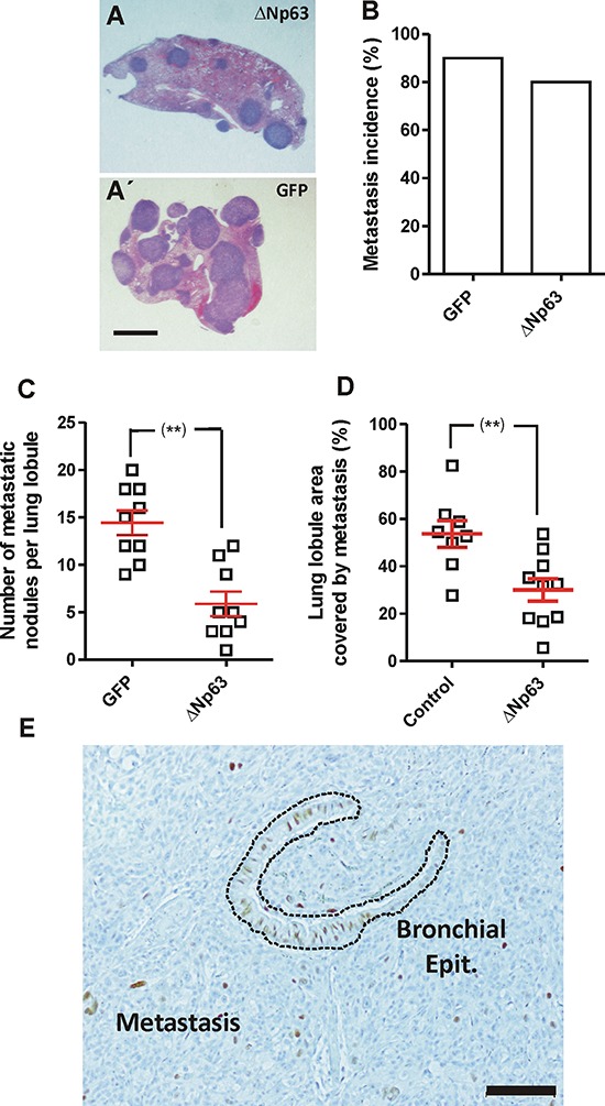
Figure 5. Expression of huΔNp63α decreases experimental metastasis development.

A, A'. Representative images of H&E stained lung lobule sections showing the presence of metastatic nodules upon tail vein injection of 940 cells upon transduction with ΔNp63α (A) or GFP (A') coding lentiviruses. Bar = 0.5 cm B–D. Summary of lung metastasis incidence (B), number of metastatic nodules per lung lobule (C) and lung area covered by metastatic nodules (D), observed in mice injected into the tail vein with 940 or huΔNp63α cells and sacrificed one month after injection. **denotes a p value < 0.01 E. Representative immunohistochemistry image showing p63 expression in a lung metastasis produced by the tail vein injection of huΔNp63α 940 cells. Bar = 250 μm.
