
Fig. 6.
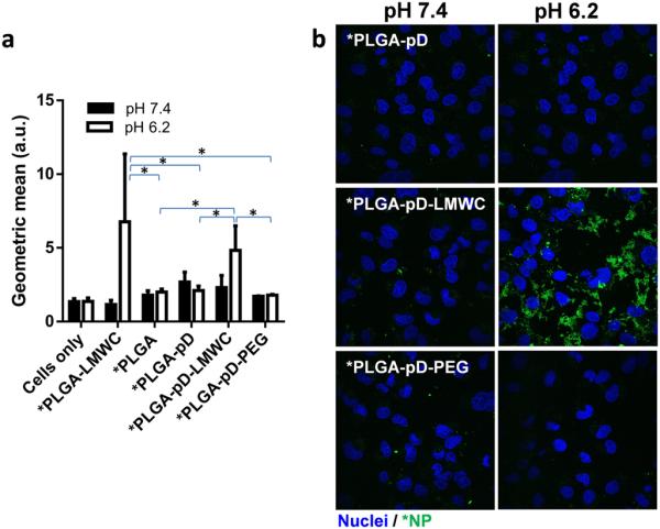
pH dependent interaction of *PLGA-pD, *PLGA-pD-LMWC, and *PLGA-pD-PEG NPs with SKOV-3 cells, (a) quantified by Flow-cytometry (Geometric mean at pH 6.2 was significantly different for *PLGA-LMWC and *PLGA-pD-LMWC from that of *PLGA, *PLGA-pD and *PLGA-pD-PEG, *: p <0.05 by two tailed t-test) and (b) visualized via confocal microscopy, after 3 hours of incubation (Green: *NPs; blue: nuclei).