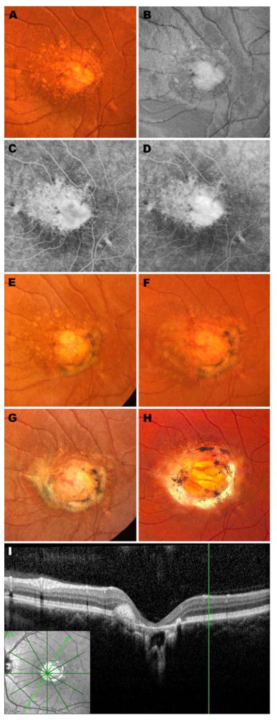
Figure 1.
Retinal images spanning 30 years from the left eye of an affected member of Family A: color fundus photograph (A), red free fundus photograph (B), early phase fluorescein angiogram (C) and late phase fluorescein angiogram (D) at age 6; color fundus photographs at ages 8 (E), 10 (F), 11 (G) and 33 years (H); optical coherence tomogram at age 33 years (I, J). This patient has been previously reported (Table 1). Stereo images of panels B–D are provided in Supplemental Figure 2 (available at http://aaojournal.org).

