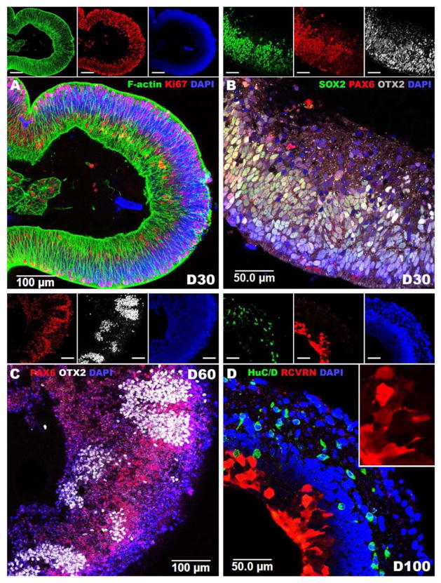
Figure 3.
Using normal human iPSCs to model retinal development. A–D: Immunocytochemical analysis of iPSC derived eyecup-like structures targeted against F-actin (Phalloidin), SOX2, PAX6, OTX2, HuC/D and recoverin (RCVRN). After 30 days of differentiation (D30) polarized neural epithelia (A, F-Actin - green) comprised of proliferating cells (A, Ki67 - red) positive for the early retinal progenitor cell markers SOX2 (B, green), PAX6 (B, red) and OTX2 (B, white) are present. After 60 days of differentiation, PAX6 (C, red) expression is restricted to OTX2 negative presumptive RPE while OTX2 (C, white) is restricted to PAX6 negative photoreceptor precursor cells. After 100 days of differentiation, eyecups are laminated with HuC/D-positive (D, green) ganglion cell like neurons in the inner layer and recoverin-positive (D, Red) photoreceptor precursor cells in the outer layer. Insets depict individual fluorescent channels. A & D: 40X magnification. B & C: 20X magnification.

