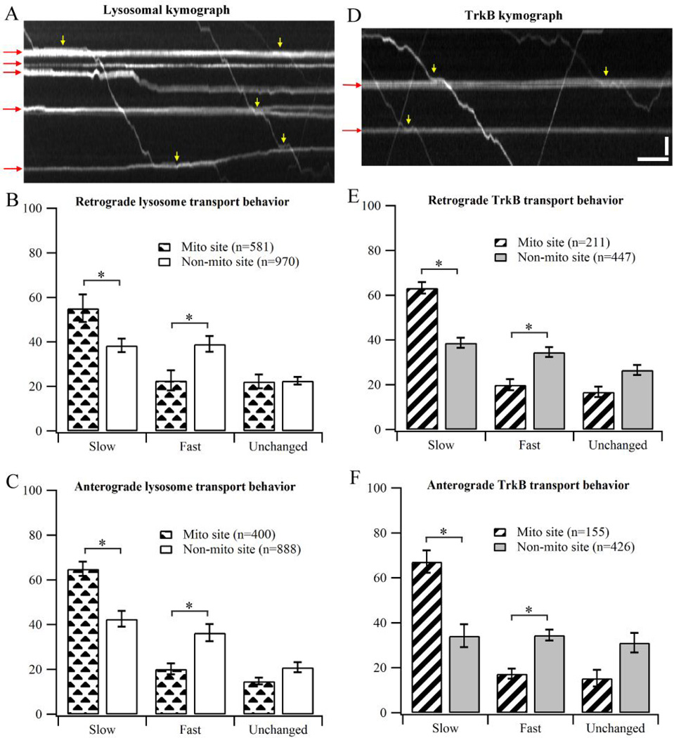
Figure 3.
Transport behavior of lysosomes and TrkB when crossing stationary mitochondria. (A) Representative kymograph showing transport behavior of lysosomes in DRG neurons. Red arrows indicate stationary mitochondrial traces in the kymograph. Yellow arrows show the regions where the cargos clearly slow down at mitochondrial sites. (B–C) Transport behavior of lysosomes in DRG neurons. Data were cumulated from 3 independent cultures. (D) Representative kymograph showing transport behavior of TrkB in hippocampal neurons. (E–F) Behavior of TrkB transport in hippocampal neurons. Data were cumulated from 4 independent cultures. Error bars show standard deviation. Bootstrapping (10 sets of 200 randomly selected events) was performed to calculate the standard deviation. Statistical significance was assessed using Student’s t-test, p<0.001. Horizontal bar, 5s.Vertical bar, 5 µm.

