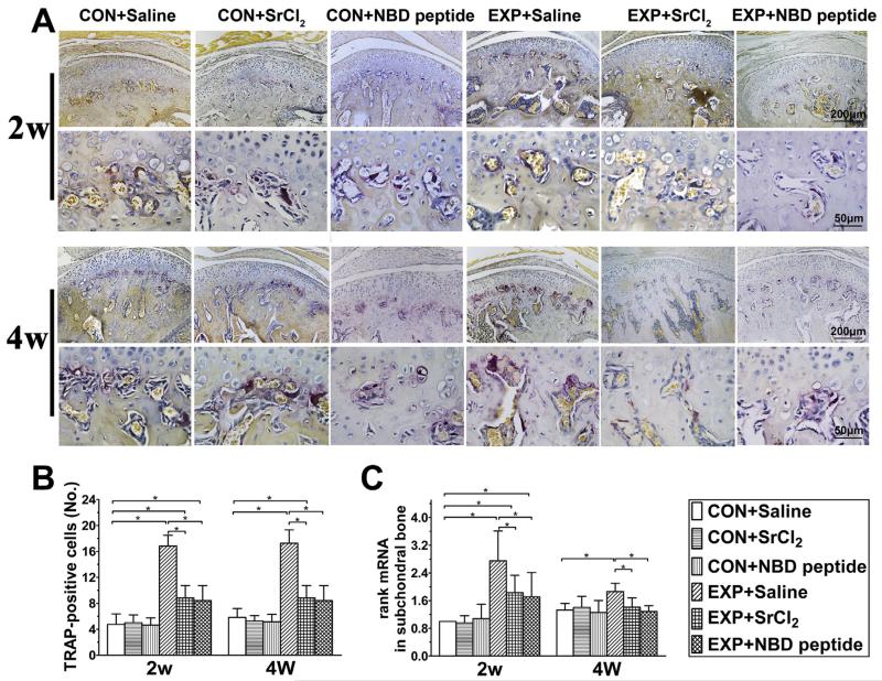
Fig. 6.
The osteoclast numbers in the mandibular condylar subchondral bone in the control and UAC mice. (A) TRAP staining in the mouse mandibular condylar subchondral bone. Bars = 200 μm in the low-magnification images and Bars = 50 μm in the high-magnification images. (B) Quantitative analysis of TRAP-positive osteoclasts (n = 6). P values: 2w: P-values between groups with * are less than 0.001; 4w: CON + Saline vs UAC + Saline and UAC + SrCl2: P < 0.001 and P = 0.032; UAC + Saline vs UAC + SrCl2 and UAC + NBD peptide: both P < 0.001. (C) mRNA expressing levels of rank in condylar subchondral bone were shown (n = 3). P values: 2w: CON + Saline vs UAC + Saline, UAC + SrCl2 and UAC + NBD peptide: P < 0.001, P = 0.005 and P = 0.015; UAC + Saline vs UAC + SrCl2 and UAC + NBD peptide: P = 0.002 and P = 0.001; 4w: CON + Saline vs UAC + Saline: P = 0.001; UAC + Saline vs UAC + SrCl2 and UAC + NBD peptide: P = 0.002 and P < 0.001. Data are expressed as means and 95% confidence intervals (CIs); * indicates statistically significant differences between groups.

