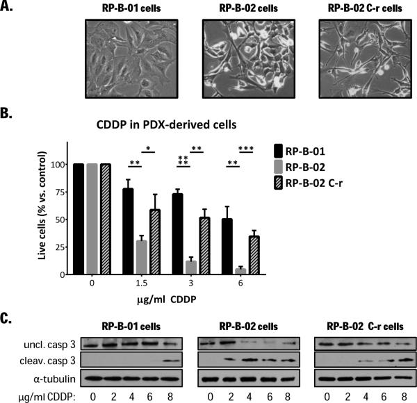
Figure 2. PDX-derived UCC cells retain the same cisplatin sensitivity displayed in vivo.
A. Bright field pictures of RP-B-01, RP-B-02 and RP-B-02 C-r cells isolated from PDXs. B. Propidium iodide exclusion assay on PDX-derived UCC cancer cells, treated with increasing concentrations of CDDP for 48 hours (mean ± SD). C. Western blot analysis of uncleaved and cleaved caspase 3 in PDX-derived UCC cells treated as described in panel B. α-tubulin served as loading control. *p <0.05, **p <0.01, ***p <0.001, ****p <0.0001,as compared to the same CDDP concentration in RP-B-02, using two tailed t-test analysis.

