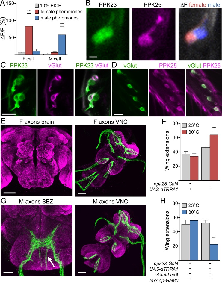
Figure 1. F and M cells comprise distinct chemosensory neuron classes.
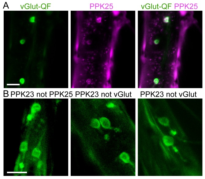
(A) F cells (PPK23+ PPK25+) respond to female pheromones whereas M cells (PPK23+ PPK25-) respond to male pheromones; n = 7 bristles. The female pheromone mix contained 7,11-heptacosadiene and 7,11-nonacosadiene. The male pheromone mix contained 7-tricosene and cis-vaccenyl acetate. (B) GCaMP6s marks two PPK23 cells per bristle (left). CD8::tdTomato marks the PPK25 cell (middle). Maximum ΔF of both PPK23 cells to female (red) or male pheromones (blue) (right). (C) vGlut (magenta) is expressed in one PPK23 cell (green) under each bristle. Transgenic flies with ppk23-LexA, lexAop-CD2::GFP, vGlut-Gal4, UAS-CD8::tdTomato were used for cell labeling. (D) vGlut (green) and PPK25 (magenta) are expressed in the same cell under each bristle. Flies contained vGlut-LexA::VP16, lexAop-CD2::GFP, ppk25-Gal4, UAS-CD8::tdTomato. (E) Axons from F cells in the legs (green) do not project to the central brain (left) but instead terminate in the six leg neuromeres of the ventral nerve cord (VNC, right). Brains are counterstained with nc82 (magenta) to show neuropil. (F) Expression of dTRPA1 in F cells promoted male-female courtship upon heat-evoked neural activation; n = 11–21/condition. Number of unilateral wing extensions per 10-minute trial was recorded. (G) M cells from the legs project to the SEZ in the brain (arrow shows fibers entering from the cervical connective) and VNC. Other SEZ axons come from the proboscis, with fibers entering from the labellar nerve. (H) dTRPA1-mediated activation of M cells suppressed male-female courtship. n = 10–12/condition. Number of unilateral wing extensions per 10-minute trial was recorded. Scale bars, 5 (B), 10, (C, D) 25 (G, SEZ) or 50 μm (E, G, VNC). Data are Mean ± SEM. Kruskal-Wallis test, Dunn’s post-hoc (A) or 2-way ANOVA, Bonferroni post-hoc (F, H). **p< 0.01. See also Figure 1—figure supplement 1, on selectively targeting F or M cells.


