. 2015 Oct 14;4:e05360. doi: 10.7554/eLife.05360

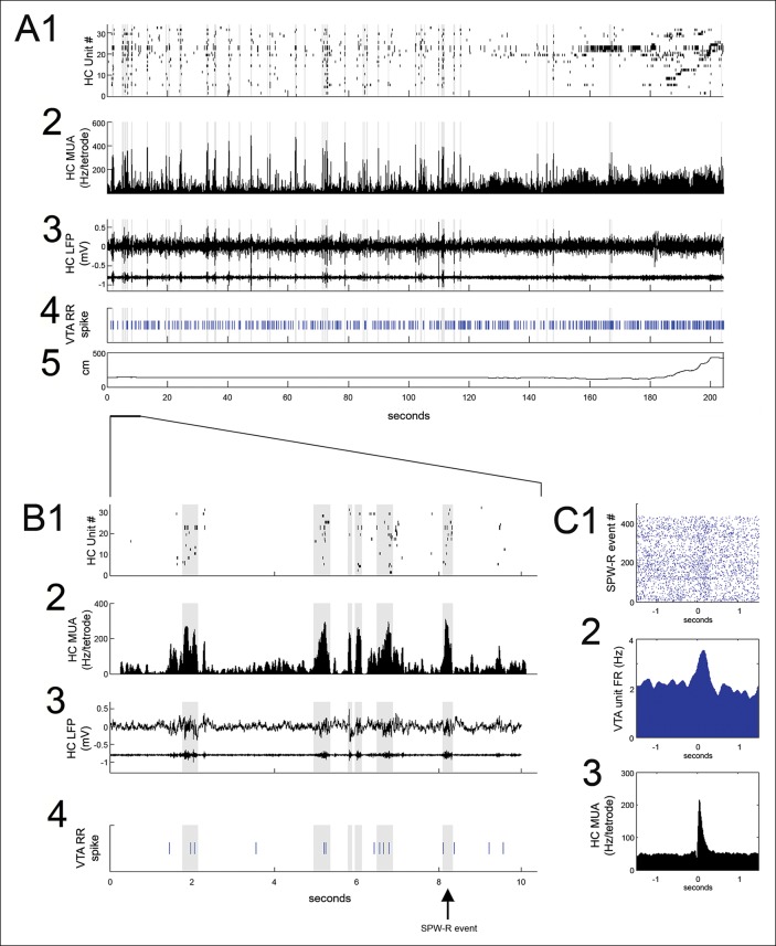
Figure 2. VTA unit coordination with hippocampal sharp-wave ripples.

(A) Continuous recordings of hippocampal (HC) (1) single unit activity, (2) multiunit activity (MUA, average spike rate per tetrode), (3) local field potential and ripple band, (4) a simultaneously recorded reward-responsive (RR) VTA unit, and (5) the animal’s position on the track. The hippocampal units are ordered by the position of their place fields on the spatial working memory task. Sharp-wave ripple events (SPW-R) are shown in gray. (B) A magnified view of 10 s of continuous data. (C1) Rastered RR VTA unit action potentials, (2) RR VTA unit peri-event time histogram (PETH; smoothing with a 50ms Gaussian window), and (3) HC multiunit PETH (10 ms Gaussian smoothing), aligned to the start of SPW-R-associated HC multiunit events.

DOI: http://dx.doi.org/10.7554/eLife.05360.004

Figure 2.

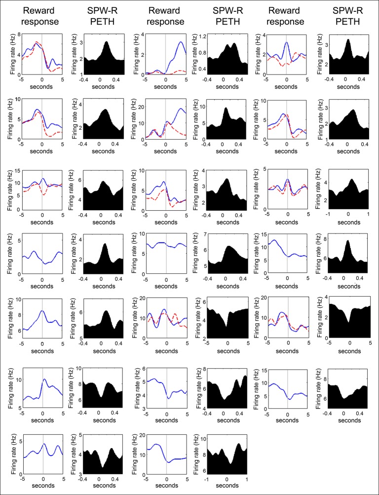
Figure 2—figure supplement 1. Firing rate distributions of SPW-R modulated VTA units at reward acquisition and at SPW-R events of quiet wakefulness.

Figure 2—figure supplement 1.

For units recorded on the SWM task, the average nosepoke triggered PETH for correct trials (solid blue lines) and for error trials (red dashed lines) are shown. Units acquired on the linear maze have a single nosepoke triggered PETH. Data are aligned to the time of nosepoke (vertical line). For the SPW-R event triggered PETH plots, data are aligned to the start of SPW-R events. Note that VTA unit activity often increases during reward approach and reward acquisition, and that VTA unit activity can be both positively and negatively modulated at SPW-R events.