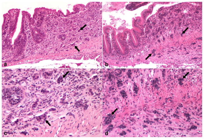
Fig. 2.
Danio rerio, intestine with neoplasia. (a) Fish 1. Intestinal adenocarcinoma with neoplastic cells (arrows) within the lamina propria and muscularis, extending to the serosal layer. HE. (b) Fish 7. Intestinal small cell carcinoma infiltrating the lamina propria with invasion into the muscularis (arrows). HE. (c) Fish 1. Higher magnification of the intestinal adenocarcinoma in (a) shows neoplastic cells forming solid and pseudoacinar structures. (d) Fish 7. Higher magnification of Intestinal small cell carcinoma in (b) shows fusiform neoplastic cells forming small aggregated nests.

