. 2015 Sep 16;8(1):69–78. doi: 10.4168/aair.2016.8.1.69

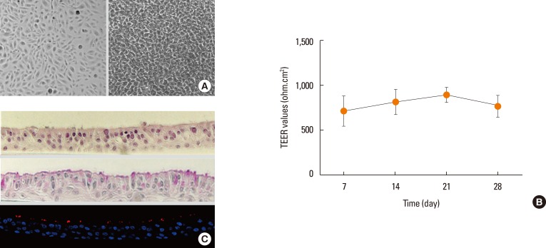
Fig. 1. Morphology of cultured nasal epithelial cells under light microscope, measurement of transepithelial electric resistance (TEER) and histologic appearance of ALI cultures of NHNE cells. (A) Primary nasal epithelial cells growing on subculture (left panel) and ALI culture periods (right panel) (original magnification ×20). (B) TEER of unstimulated NHNE cells cultured for 7, 14, 21, and 28 days. Values are expressed as mean±SD. (C) Cross section (5 µm) of NHNE cells, at 14 days after confluence, stained with H&E (upper panel), PAS (middle panel) and immunofluorescence (lower panel, red color: α-tubulin IV, blue color: DAPI) (original magnification ×200).

Fig. 1