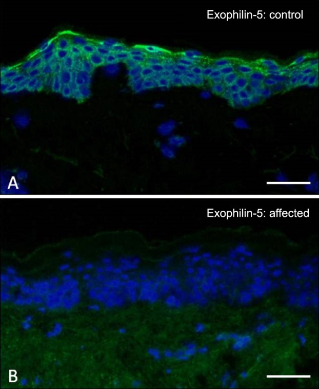
Fig. 7. Immunofluorescence microscopy labeling of skin for exophilin-5. (A) In healthy control skin, there is bright pan-epidermal cytoplasmic staining. (B) In contrast, in skin from a patient with biallelic EXPH5 mutations, there is a complete absence of immunostaining. Scale bar=50 µm.

