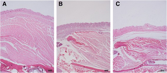
Fig. 3.

Histological view. a The saline-treated group, b 5-U BTX-treated group, c 10-U BTX-treated group. Interestingly, the thickness of the mandibular ramus (asterisk) was changed after BTX injection. Degenerative change was also shown in both the 5- and 10-U BTX-treated groups (hematoxylin and eosin stain, original magnification ×20)
