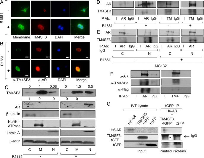
Figure 4.
TM4SF3 colocalizes and interacts with AR in PCa cells. LNCaP cells were treated with either ethanol (−) or 1nM R1881 (+) for 24 hours and measured for expression of TM4SF3 and AR by (A and B) immunocytochemistry or (C) Western blotting of cell fractions cytosol (C), membrane (M), and nuclear (N) using antibodies against TM4SF3, AR, and β-actin, which was used as loading control. Note that that the membrane marker (CellMask from Life Technologies) in A stains the plasma membrane, and DAPI was used to stain the nuclei in both A and B. Scale bar in B, is 10 μm. Cell fractions markers are β-tubulin (cytosol), Na+/K+ ATPase (membrane), and Lamin A (nuclear). LNCaP cells were treated with ethanol (−) or 1nM R1881 (+) for 24 hours and (D) whole-cell extracts or (E) cytosolic (C) and nuclear (N) extracts were subjected to IP using antibodies against AR or TM4SF3 or IgG. Note that cells in E were also treated with 10μM MG132. F, TM4SF3-Flag and AR or (G) TM4SF3-tGFP, tGFP, and H6-AR were expressed in vitro using TNT system and (F) mixed and subjected to IP using anti-AR or anti-TM4SF3 antibody or (G) Cobalt pull-down purified H6-AR was added to IP-purified TM4SF3-tGFP or tGFP. Western blotting was used to measure levels of AR, H6-AR, TM4SF3, TM4SF3-Flag, and TM4SF3-tGFP, and tGFP in C–G, including an anti-Flag antibody in F. In tGFP IP of G; **, TM4SF3-tGFP and *, IgG heavy chain. For C, the numbers above the gel represent average quantifications of 3 TM4SF3 and AR Western blottings, standardized to β-actin, and relative to the first lane, which was set to 1.

