Figure 5.
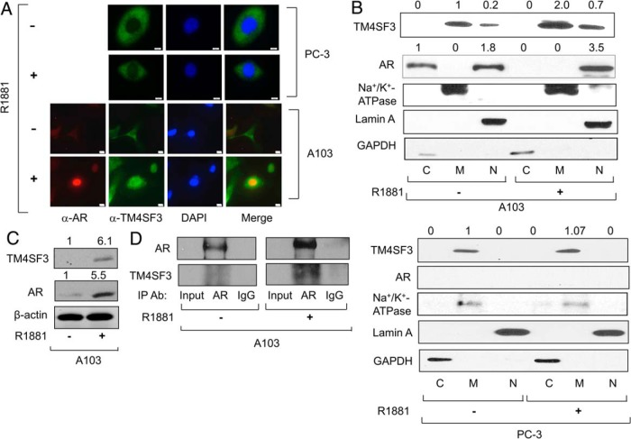
TM4SF3 colocalizes and interacts with AR in AR-expressing PC-3 cells. PC-3 or A103 cells were treated with either ethanol (−) or 1nM R1881 (+) and measured for expression of TM4SF3 and AR by (A) immunocytochemistry or (B and C) Western blotting of (B) A103 or PC-3 cell fractions (cytosol [C], membrane [M], and nuclear [N]) or of (C) A103 whole-cell extracts. Scale bar in A, 10μM. Cell fractions markers are GAPDH (cytosol) marker, Na+/K+ ATPase (membrane), and Lamin A (nuclear marker); β-actin controls for protein loading. D, IP experiments were performed with whole-cells extracts from A103 cells treated with ethanol (−) or 1nM R1881 (+) using antibodies against AR or IgG. Western blotting was used to measure levels of copurified AR and TM4SF3. DAPI was used to stain the nuclei in A. For B and C, the numbers above the gel represent average quantification of 3 TM4SF3 and AR Western blottings, standardized to β-actin, and relative to the first lane, which was set to 1.

