Figure 7.
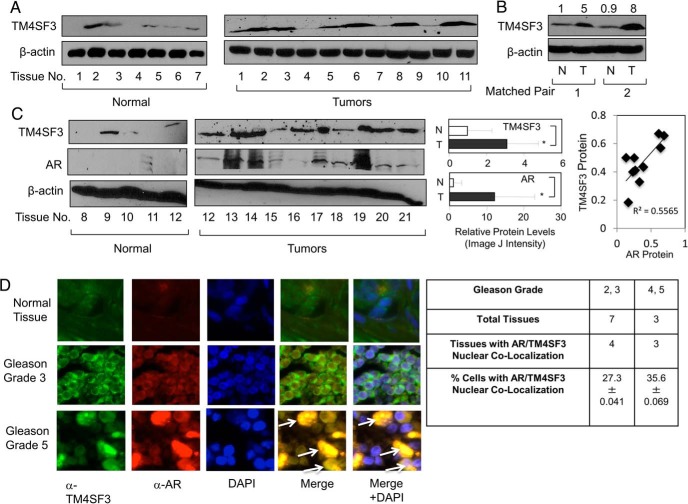
Nuclear TM4SF3 and AR are overexpressed in PCa. Western blotting was used to measure TM4SF3 and AR expression in (A and C) normal and tumor tissues and (B) matched pairs of normal (N) and tumor (T) from same patients. Note that β-actin was used as a loading control. TM4SF3 and AR protein levels were significantly higher (P < .05) in tumors as compared with normal, analyzed by Student's t test. Linear regression analysis was done to compare protein levels of TM4SF3 and AR, which were significant as represented by R2 = 0.55655 and P = .03. It is important to mention that both normal prostate and cancer tissues have been previously shown (see Ref. 6) to have mainly epithelial cells based, on expression of cytokeratin 18 (K18), a maker of epithelial cells, and keratinocyte growth factor (KGF), a marker gene for prostate stromal cells. For B, the numbers above the gel represent quantification of the TM4SF3 Western blotting signals, standardized to β-actin, and relative to the first lane, which was set to 1. D, Normal prostate tissue and different grades of prostate tumors (from US Biomax) were subjected to immunocytochemistry to monitor the relative expression levels and subcellular localization TM4SF3 and AR. DAPI stains nuclei. Arrows in merge images shows colocalization of TM4SF3 and AR in both nucleus and cytoplasm. Table shows quantification of number of tumors and % of cells having positive AR/TM4SF3 nuclear colocalization (±SD). Student's t test showed that a statistically significant higher (P < .05) number of cells of grade 4/5 than grade 2/3.

