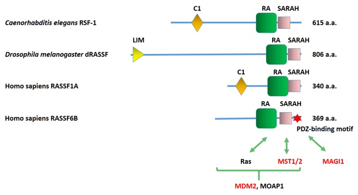
Figure 1.
Structures of Caernorhabditis elegans RSF-1, Drosophila melanogaster dRASSF. Homo sapiens RASSF1A, and Homo sapiens RASSF6. C1, phorbol esters/diacylglycerol-binding domain. RA, Ras association domain. SARAH, Salvador/RASSF/Hippo domain. LIM, Zinc-binding domain present in Lin-11, Isl-1, Mec-3. The PDZ-binding motif of RASSF6 is depicted by a red star. The amino acid number of each protein is shown on the right. The RASSF6-interacting proteins are shown on the bottom. The interactions with MST1/2. MAGI1, and MDM2 are demonstrated at the endogenous level (red letters). Ras binds to the RA domain. MST1/2 (mammalian Ste20-like kinase 1/2) interacts with the SARAH domain. MAGI1 (membrane-associated guanylate kinase inverted 1) binds to the PDZ-binding motif. The interacting regions of MDM2 and MOAP1 (modulator apoptosis 1) are not precisely determined.

