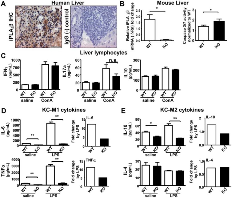
Figure 2.
iPLA2β deficiency leads to increased hepatic apoptosis associated with suppressed M1 cytokine release by KC either spontaneously or during LPS-stimulation. Male mice at 3 months old were used. Liver lymphocytes were treated with 10 µg/mL ConA for 48 h. KC were treated with 1 µg/mL LPS for 7 h. (A) iPLA2β IHC of human liver showed positive brown staining. IgG was used as (-) control; (B) In left panel, iPLA2β mRNA expression in livers of young mice was determined by qRT-PCR. In right panel, caspase 3/7 activity measured by luminescence normalized to the WT levels was obtained in liver homogenates of WT and KO mice (N = 3 per group for PCR and N = 4–5 per group for luminescence); (C) Spontaneous or ConA-stimulated release of IFNγ, IL-17 and IL-10 measured by ELISA was determined in liver lymphocytes of 3-month old WT and KO (N = 4–6 per group); Spontaneous or LPS-stimulated release of IL-6 and TNFα (D) as well as IL-10 and Il-4 (E) measured by ELISA was determined in KC isolated from WT and KO (left panel), and their fold increase by LPS was calculated (right panel) (N = 6 per group). * p < 0.05 vs. WT; ** p < 0.005 vs. WT.

