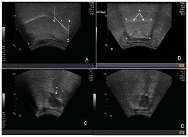
Fig. 1.
Transcervical ultrasound examination of oropharynx. Parasagittal view (A) demonstrates intact hyperechoic oral mucosa of oral and base of tongue (+). The hyoid (ψ) and mandibular (*) shadows are shown. The sonographic base of tongue is the posterior one third of the distance between the central portions of the mandible and hyoid annotated by (↓). The palatine tonsillar region is isoechoic (Φ). On coronal view (B) hyoid shadows are seen bilaterally (ψ). The oral mucosa is intact (+). The base of tongue musculature is noted to be symmetric. Panels C and D are images representative of a base of tongue lesion. On parasagittal view is a hypoechoic lesion, the majority of which is beneath the mucosal surface of the base of tongue, with a slight disruption of the mucosa (xx). The mass is also visualized in coronal view (D). Extent across midline is appreciated in coronal view.

