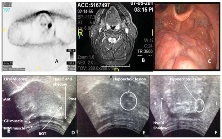
Fig. 2.
Clinical and radiographic images of a patient with head and neck squamous cell cancer of unknown primary. PET scan (A), MRI (B) and fiberoptic laryngoscopy (C) do not demonstrate any evidence of a primary lesion in the oropharynx. On transcervical ultrasound, right parasagittal view (D) posterior and anterior are labeled “post” and “ant”. Normal base of tongue (BOT) without evidence of a tonsillar mass is shown in panel D. The mucosa of the oral and base of tongue is intact. The myelohoid (MM) and geniohyoid (GH) muscles are visualized and intact. On left parasagittal view (E) a hypoechoic lesion, relative to the isoechoic normal tongue is appreciated and is consistent with a suspected base of tongue mass. This is confirmed on coronal view (F), whereby a small hypoechoic lesion is similarly appreciated.

