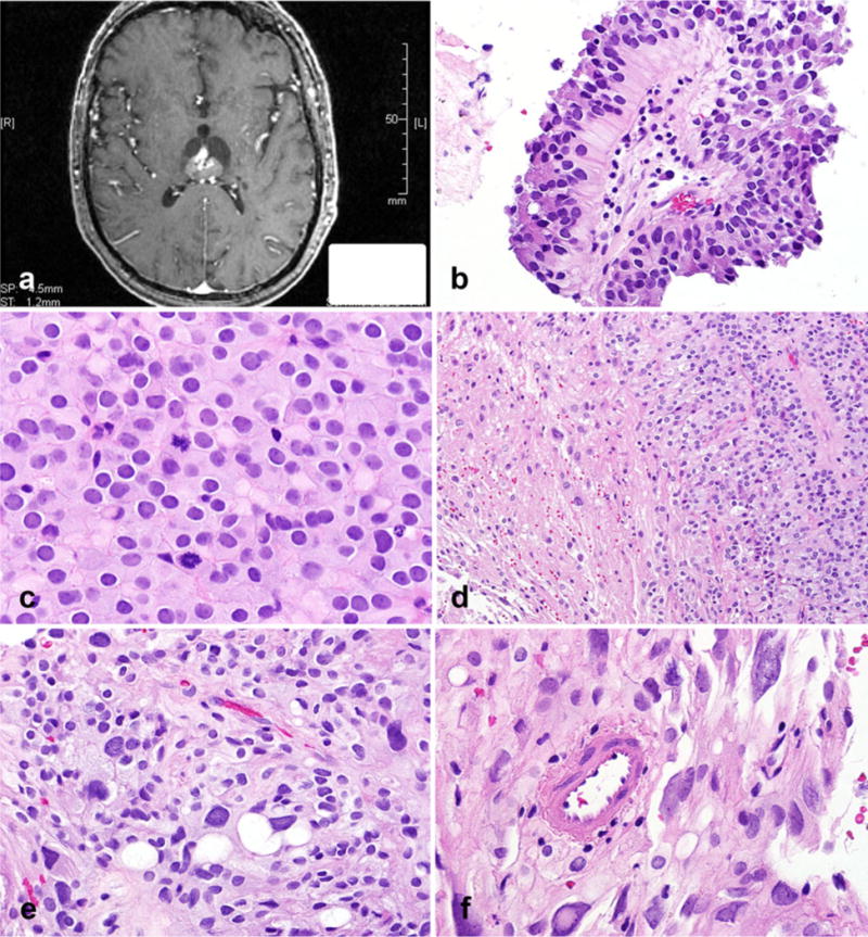
Fig. 1.

Papillary tumor of the pineal region with biphasic areas. Heterogeneously enhancing, partially cystic lesion within the dorsal aspect of the third ventricle (axial T1-weighted image, post-contrast) (a). Focal areas composed of pseudopapillae were a feature (b) as also were sheets of tumor cells with a vague perivascular arrangement, cellular monotony, and increased mitotic activity (c). Sharp interface between classic papillary areas and distinct pleomorphic cells (d). Vacuoles of different sizes in pleomorphic areas (e). Pleomorphic areas with cells containing glassy, poorly defined cytoplasm, nuclear pleomorphism, intranuclear inclusions with hyperchromasia, and smudged chromatin (f)
