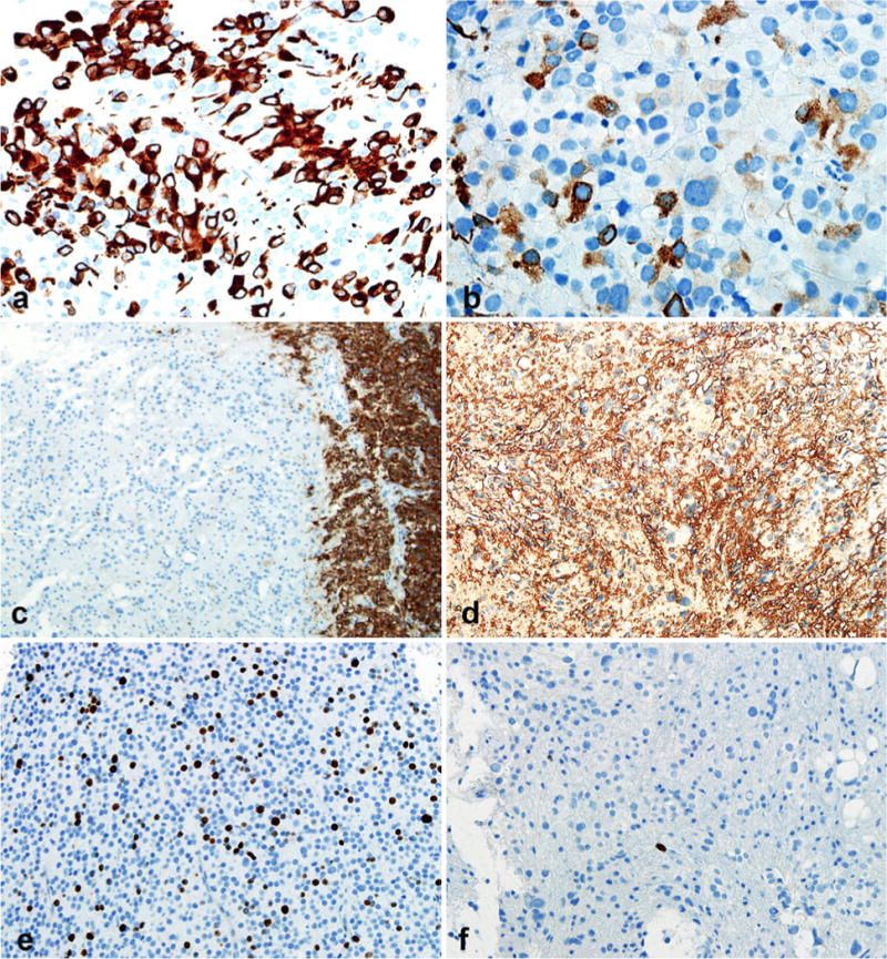
Fig. 2.

Contrasting immunohistochemical features in biphasic areas. Papillary areas with strong cytokeratin (CAM 5.2) expression (a). The strength of CAM 5.2 labeling was weaker in pleomorphic areas (b). Synaptophysin was not expressed in pleomorphic areas (left) in contrast with adjacent pineal gland (right) (c), whereas strong S-100 expression was present in all areas (d). Paradoxical MIB1 labeling indices were present, being elevated in cellular monotonous areas (e) and extremely low in pleomorphic areas (f)
