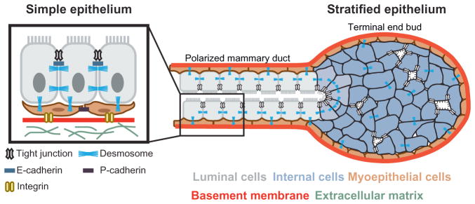
Figure 1.
Normal transitions in adhesion during epithelial branching morphogenesis. Mammary epithelium initiates branching morphogenesis postnatally. Tube elongation is accomplished by a stratified terminal end bud, which contains many internal luminal cells that lack apicobasal polarity and display reduced numbers of intercellular junctions. The epithelium at the rear polarizes to a bilayered, simple ductal architecture consisting of an inner layer of luminal cells and a basal layer of myoepithelial cells. Epithelial cells in the ducts are connected by many cell–cell junctions. Schematic adapted from original by Robert Huebner, with permission.

