Fig. 1.

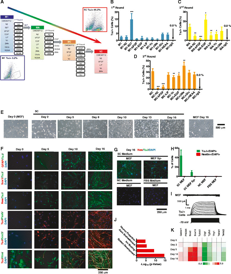
Development of 5C medium that directly converts MEFs to cells with neuronal characteristics. a Schematic illustration of the steps taken to develop the 5C medium (conversion efficiency as high as 46.3 %) from MC, MA, and the original M1 medium (efficiency around 3.2 %) during the first three rounds of factor deduction. b–d Percentages of TuJ+ cells determined by FACS during four rounds of factor reduction. The components of all medium were listed in Additional file 1: Table S1. Antibody against IgG was used as a negative control during FACS analysis. e Morphologies of cells on days 0, 2, 5, 8, 10, 13, and 16 during reprogramming were provided as phase-contrast images. f GFAP, Sox1, Nestin (Nes), Pax6, Sox2, or Vim were stained together with TuJ and DAPI on days 0, 5, 10, and 16 during reprogramming. g–h MEFs generated without the spinal cord (sp-), normal MEFs were cultured with 5C medium for 16 days and subjected for TuJ and Nestin staining. Another two groups of MEFs were cultured with NC medium and FBS medium, respectively. Nestin, TuJ, and DAPI were stained on day 16 (g). Percentage of Nestin+ and TuJ+ cells was summarized in h. i Representative recordings of voltage-gated ion channels from a TuJ+ cells. j–k RNA-seq were performed on days 0, 2, 5, 10, and 16 during 5C-induced trans-differentiation. Genes with significant up-regulation (over twofold) were used for Gene Ontology analysis. The top four enriched clusters were listed in j. The expression of five neuron markers and five fibroblast markers from RNA-seq was listed in k
