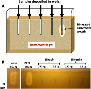
Fig. 1.

Heme detection setup and specificity of porphyrin detection. a. A vertical gel was set up with a 0.8 cm spacer. The gel comprised 0.6 % agarose and growth medium; Bacteroides (~105 cfu/ml) was resuspended in medium, which was then poured in the pre-warmed apparatus. Metal prongs (5 cm) were used to form the wells. Samples were loaded in wells, and gels were overlaid with a 5 ml, 1.2 % agar plug. Bacteroides growth stimulation was visualized as dense growth around wells after overnight incubation at 37 °C, as schematized for the rightmost well. b. Heme and PPIX, but not heme breakdown products stimulated B. thetaiotaomicron (Bt) growth. The indicated amounts of the tested products were loaded in wells of the gel setup described in "A". A ~0.5 to 1 cm no-growth-zone from the top of the gel was due to aerobic inhibition of Bacteroides growth
