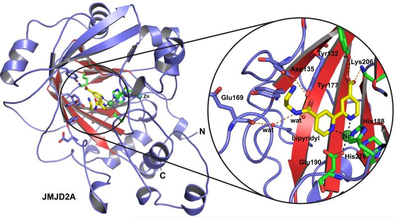
Figure 3.
Views from the crystal structure of JMJD2A in complex with compound 13a (yellow sticks). The double-stranded β-helix (conserved in 2OG oxygenases) is in red. Residues that bind Fe(II) and 2OG are shown as green sticks, Ni(II), which replaces Fe(II) for crystallography, is shown as a green sphere. Other residues likely interacting with 13a are shown as blue sticks.

