
Fig. 3.
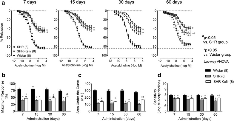
Time-course of the effects of kefir administration on the endothelial dysfunction of SHR. Dose–response curves to acetylcholine-induced relaxations of aortic rings from SHR-kefir compared to the non-treated SHR and to the normotensive Wistar rats (a). The bar graphs show the maximum relaxation (b), the area under the curve (c) and the sensitivity (pEC50, d) to acetylcholine. The values shown are the mean ± SEM. *p < 0.05 vs. Wistar group; #p < 0.05 vs. SHR (two-way ANOVA)